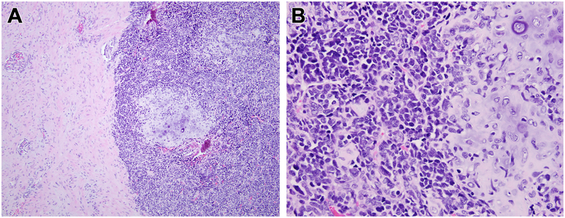
Figure 5. Histologic appearance of mesenchymal chondrosarcoma metastatic to the kidney for comparison.
At low power, one can appreciate the primitive small cells with associated nodules of cartilage to the right and entrapped kidney to the left (A). At high power (B), one can appreciate the cartilage and primitive small blue round cell appearance, which contrasts with the monomorphic spindle cell appearance of the MEIS1-NCOA2 renal sarcomas. This mesenchymal chondrosarcoma showed a NCOA2 gene rearrangement, but lacked evidence of MEIS1 gene abnormalities.

