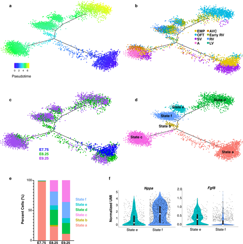
Extended Data Figure 6: Pseudotemporal ordering of myocardium populations.
Pseudotime trajectory of myocardium populations colored by a, pseudotime value, b, cluster identity, c, embryonic stage of collection and d, cell state. Pseudotime trajectory analysis was applied to n=6,474 cells. e, Percentage of cells in each state that were captured at E7.75, E8.25 or E9.25. f, Violin plots showing expression of Nppa and Fgf8 in State e and State f from pseudotime trajectory in (d). Statistics for differential gene expression tests were applied to n = 455 cells from each state. Bonferroni correction adjusted p-value < 1×10⁻⁴ (Wilcoxon rank sum test, two-sided). Summary statistics reported in violin plots: the center white line represents median gene expression and the central black rectangle spans the first quartile to the third quartile of the data distribution. The whiskers above or below the box indicate value at 1.5x interquartile range above the third quartile or below the first quartile.

