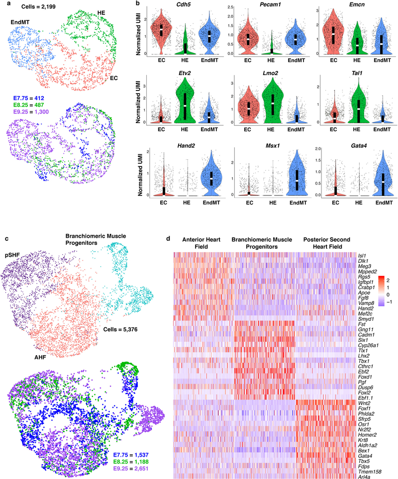
Extended Data Figure 4: Heterogeneity in endocardium/endothelium and multipotent progenitor populations.
a, UMAP plot of reclustered Endocardium/Endothelium population colored by cluster and embryonic stage of collection. b, Violin plot of markers indicating distinct subpopulations of endocardium/endothelial cells. Summary statistics reported in violin plots: the center white line represents median gene expression and the central black rectangle spans the first quartile to the third quartile of the data distribution. The whiskers above or below the box indicate value at 1.5x interquartile range above the third quartile or below the first quartile. Statistics for differential gene expression tests were applied to n=2,199 cells. c, UMAP plot of reclustered multipotent progenitor populations colored by cluster and embryonic stage of collection. d, Heatmap showing curated list of marker genes that identify pSHF, AHF and branchiomeric muscle progenitors. Scale indicates Z-scored expression values. Statistics for differential gene expression tests were applied to n=5,376 cells. HE, hemato- endothelial progenitors; EC, endocardial/endothelial cells; EndMT, endothelial-mesenchymal transition cells; AHF, anterior heart field; pSHF, posterior second heart field.

