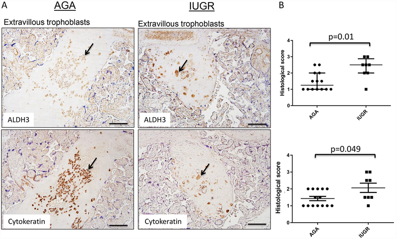
Figure 6. ALDH3 expression in extravillous trophoblasts.
Term human placentas were stained for ALDH3 expression, and representative sections of extravillous trophoblasts pictured here. Scale bars represent 100 μm. Arrows indicate positively stained cells (brown). (A) ALDH3 expression is detected exclusively in extravillous trophoblast, especially those found in close proximity to maternal decidual blood vessels. The presence of these cells was confirmed by cytokeratin staining. (B) Positivity of ALDH3 staining in extravillous trophoblast was increased in IUGR placentas scored by two independent pathologists. (PS: AGA: median=1.4, interquartile range 1.0–2.0, n=14; IUGR: median=2.1, interquartile range 1.5–2.9, n=8; p=0.01 by Mann-Whitney U testing); (BC: AGA: median: 1.0; interquartile range 1.0–2, n=14; IUGR: median=2.5, interquartile range 1.0–2.5, n=8; p=0.049 by Mann-Whitney U testing). Data are represented in scatter plots with horizontal lines representing median and interquartile range values.

