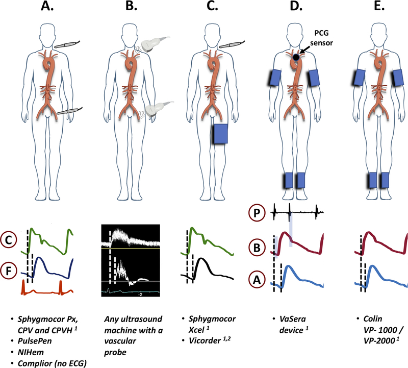
Figure 10. Methods of measurement of carotid-femoral PWV (A–C), CAVI (D) and ba-PWV (E) by various devices.
Sensors and examples of recorded signals are shown. C=carotid signal; F=femoral signal; P=phonocardiographic signal; B=brachial signal; A=ankle signal. 1: FDA-approved; 2: the vicorder uses a neck cuff, rather than a tonometry sensor.

