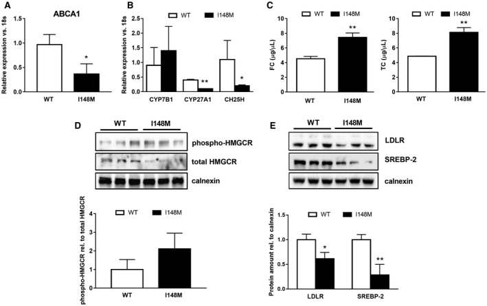
Figure 2.

Decreased LXR signaling disrupts cholesterol homeostasis in PNPLA3 I148M carrying human HSCs. Human primary HSCs isolated from liver resections and cultivated in vitro and LX‐2 stably overexpressing cells (n = 3 for each PNPLA3 genotype) obtained, as described in Materials and Methods. All bar graphs show mean values ± SD. (A,B) Expression of cholesterol efflux transporter ABCA1 and cholesterol‐metabolizing enzymes CYP7B1, CYP27A1, and CH25H analyzed by RT‐PCR and normalized to 18s in untreated primary HSCs carrying either the WT or the variant of PNPLA3 (n = 3 for each genotype); *P < 0.05, **P < 0.01. (C) TC and FC amounts quantified by using colorimetric quantitative assay on total lipid extracts collected from HSCs carrying either WT or I148M PNPLA3. Data shown are representative of two independent lipid extractions normalized to internal standards (Abcam); **P < 0.01. (D,E) Total protein extracts were isolated from primary HSCs carrying either WT or I148M PNPLA3 (n = 3 for each genotype) and analyzed by western blotting for SREBP‐2, LDLR, phospho‐HMGCR, and total HMGCR. Densitometry analysis was performed using ImageJ software, and data were normalized to calnexin. Data presented as protein amount relative to calnexin. Phospho‐HMGCR was normalized to total HMGCR and expressed as protein ratio. Open bars refer to PNPLA3 WT HSCs and closed bars to I148M HSCs (n = 3 for each genotype); *P < 0.05, **P < 0.01 versus WT carriers. Abbreviations: phospho‐, phosphorylated; rel., relative.
