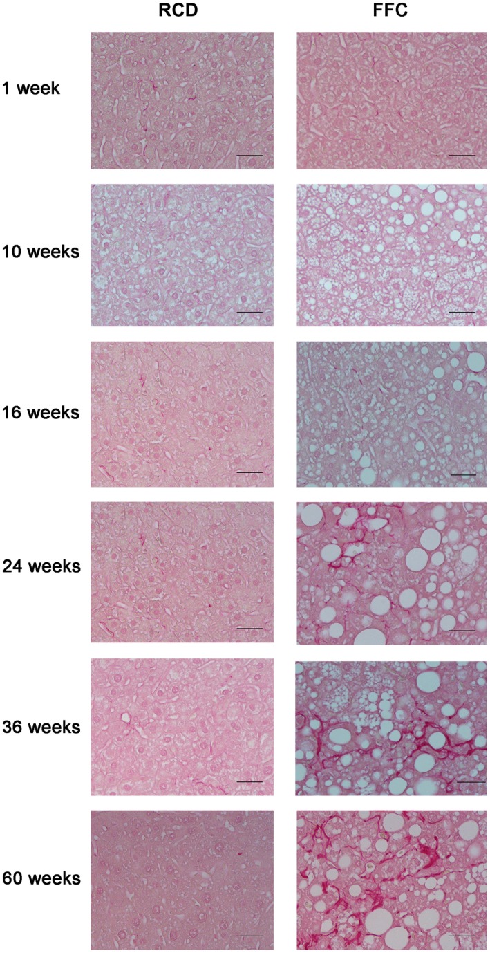
Figure 7.

Images of sirius red‐stained livers of FFC‐fed female mice. Representative images of sirius red‐stained hepatic sections (both RCD‐fed and FFC‐fed mouse samples at all time points). Original magnification ×20; n = 5‐8 per group. No fibrosis was seen in RCD‐fed mice at any time point. Most FFC‐fed mice did not show fibrosis until week 16 (fibrosis, 0; 62.5%, 5/8). From week 24 onwards, all FFC‐fed mice demonstrated progressively increased fibrosis. At week 36, all FFC‐fed mice had mild to moderate fibrosis (1a ≤ fibrosis ≤2; 100%) that persisted until the endpoint at week 60 (fibrosis, ≤2; 80%, 4/5).
