Figure 1.
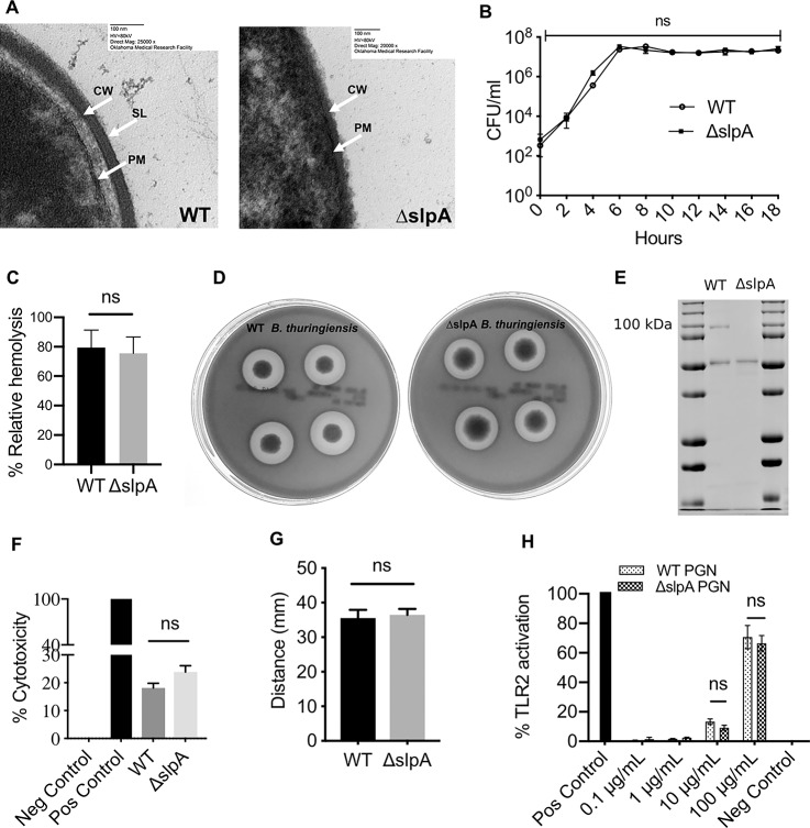
Absence of SLP does not alter bacterial growth and phenotypes. (A) Electron micrograph of thin-sections of WT and ΔslpA B. thuringiensis. CW, Cell wall; PM, plasma membrane. Magnification, ×25,000 (WT) and ×20,000 (ΔslpA). (B) In vitro growth curve of WT B. thuringiensis and its isogenic SlpA-deficient mutant (ΔslpA) in BHI broth. CFU of both WT and ΔslpA B. thuringiensis were similar at each time point (P > 0.05). Values represent the mean ± SEM for N = 3 samples per time point. (C) WT and ΔslpA B. thuringiensis were compared for their hemolytic activities (2-way ANOVA, P > 0.05). (D) WT and ΔslpA B. thuringiensis exhibited colonies with characteristic double zones of hemolysis on TSA containing 5% sheep blood. (E) Samples of the final purification step from WT and ΔslpA B. thuringiensis were loaded onto 12% polyacrylamide gel and subjected to PAGE. The 91.4-kDa band indicates the presence of SlpA in the WT B. thuringiensis, which is absent in the isogenic mutant. A 49.1-kDa flagellar protein band in both WT and ΔslpA B. thuringiensis serves as a standard. (F) Cytotoxicity of filter sterilized overnight supernatants from WT and ΔslpA B. thuringiensis in human retinal Muller cells. No significant difference was observed in the cytotoxicity of these strains (P = 0.1297). Data represents the mean ± SEM of percent of cytotoxicity for N > 5 samples. (G) The motility of WT and ΔslpA B. thuringiensis was compared. Both strains exhibited similar levels of motility (P = 0.9429). (H) Cell-wall PGN from WT and ΔslpA B. thuringiensis activated TLR2 (P = 0.8857). Data represents the mean ± SEM for N ≥ 4 samples).

