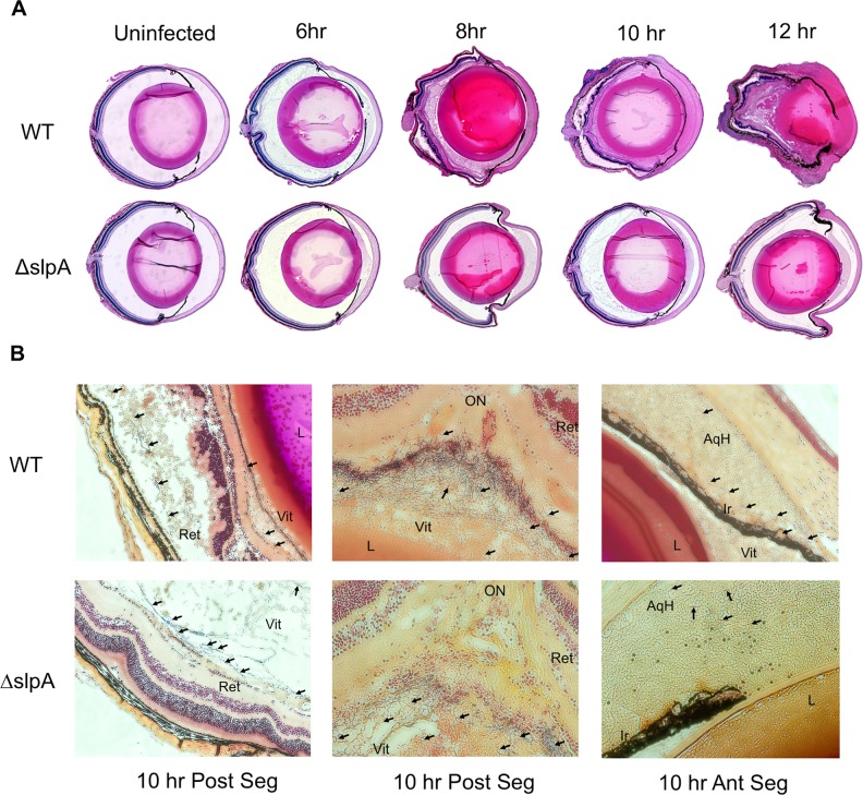
Figure 4.
Absence of SLP preserves ocular architecture in Bacillus endophthalmitis. (A) C57BL/6J mouse eyes were infected with 100 CFU of WT or ΔslpA B. thuringiensis. Infected eyes were harvested at 6, 8, 10, and 12 hours postinfection and processed for H&E and Gram staining. Magnification, ×10. (B) Bacteria were observed in the midvitreous of eyes infected with each strain at 10 hours postinfection. At this time point, WT and ΔslpA mutant were each observed in the aqueous humor of the anterior segment (Ant Seg), as well as in the midvitreous and near the retina of the posterior segment (Post Seg). Sections are representative of three eyes per time point. WT and ΔslpA B. thuringiensis are denoted by black arrows. Magnification, ×200. Ret, retina; Vit, vitreous; AqH, aqueous humor; Ir, iris; L, lens.

