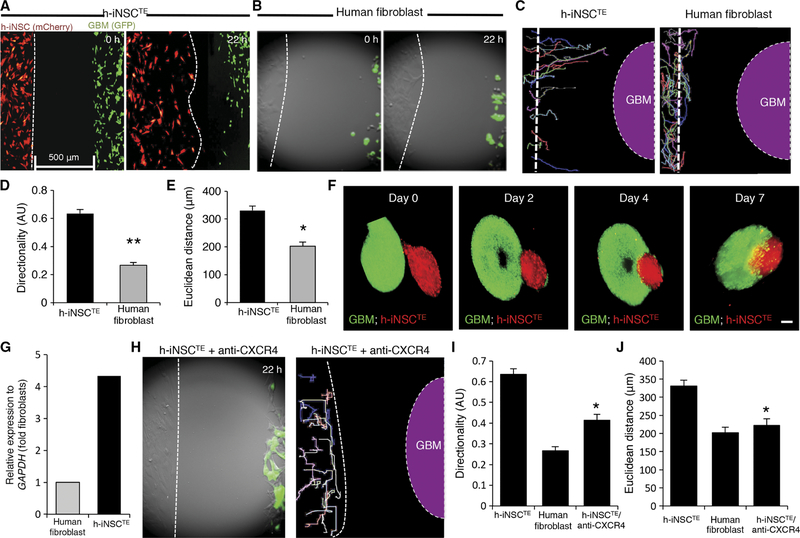
Fig. 3. Engineered h-iNSCTE homing to GBM.
h-iNSCTE–mC-FL were seeded 500 μm away from mCherry-expressing human GBM cells and placed in a fluorescence incubator microscope. Time-lapse fluorescence images were captured every 20 min for 22 hours and used to construct movies that revealed the migration of h-iNSCTE to GBM in real time. (A and B) Summary images showing migration of h-iNSCTE–mC-FL (red) (A) or parental human fibroblasts (B) toward U87–GFP-FL (green) at 0 and 22 hours after plating. (C) Single-cell tracings depicting the paths of h-iNSCTE–mC-FL or human fibroblast–directed migration toward GBM over 22 hours. Dashed line indicates the site of GBM seeding. (D and E) Summary graphs showing the directionality (D) and Euclidean distance (E) of h-iNSCTE or fibroblast migration toward GBM cells determined from the real-time motion analysis. **P = 0.00001, *P = 0.00049 by Student’s t test. (F) Fluorescence imaging showed the migration of h-iNSCTE–mC-FL (red) into U87 spheroids (green) and their penetration toward the core of the tumor spheroid over time in 3D levitation culture systems. (G) Summary graph of RT-PCR analysis showing the increased expression of CXCR4 in h-iNSCTE compared to fibroblasts. (H) Summary image and cell tracings showing the attenuated migration of h-iNSCTE after pretreatment with CXCR4-blocking antibody. (I and J) Summary graphs demonstrating a reduction in directional migration (I) (*P = 0.0000013 by Student’s t test) and Euclidean distance (J) (*P = 0.0000247 by Student’s t test) by h-iNSCTE treated with anti-CXCR4 antibodies. Data in (D), (E), (I), and (J) are means ± SEM of three independent experiments performed in triplicate. Scale bars, 200 μm.

