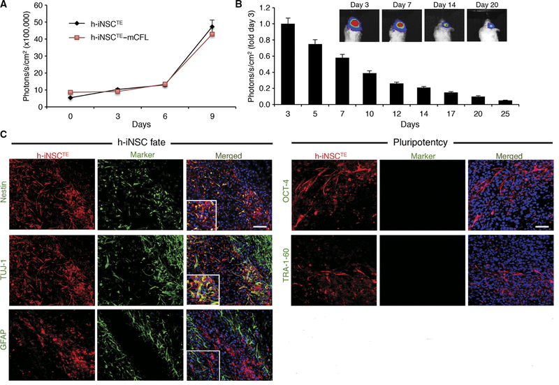
Fig. 4. In vivo characterization of h-iNSCTE transplanted into the mouse brain.
(A) Summary graph demonstrating the proliferation of unmodified h-iNSCTE and h-iNSCTE engineered to express mCherry/FL. No significant difference was found by two-way analysis of variance (ANOVA). (B and C) h-iNSCTE–mC-FL were implanted into the frontal lobes of mice, and serial BLI was used to monitor their persistence over 3 weeks. Summary graphs demonstrated that the h-iNSCTE persisted for 25 days, although they were gradually cleared (B). Immunofluorescence analysis of h-iNSCTE (red) 14 days after implantation into the brain showed nestin+ and TUJ-1+ cells, but minimal h-iNSCTE stained positive for the astrocyte maker GFAP or the pluripotency markers OCT-4 and TRA-1–60 (green) (C). Data in (A) and (B) are means ± SEM. (A) and (B) represent three different experiments performed in triplicate. Scale bars, 50 μm (C).

