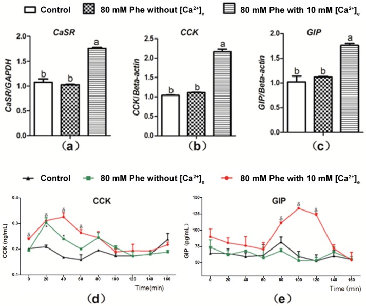
Figure 4.
Effect of [Ca2+]e on expression level of mRNA of CaSR, CCK, and GIP, as well as secretion of CCK and GIP induced by Phe in pig duodenum. Perfusion of duodenal tissues was performed with KHB without Phe and [Ca2+]e (the control group), and 80 mM Phe with or without 10 mM [Ca2+]e. After 160 min of perfusion, the tissues were collected for qPCR analysis for expression of (a) CaSR, (b) CCK, and (c) GIP; GAPDH and β-actin were used as internal controls. The perfusate solutions were obtained at a 20 min interval to determine the concentrations of (d) CCK and (e) GIP using corresponding ELISA kits. Values are presented as mean and SEM (n = 5). For each gene (a–c), means with dissimilar letters differ significantly at p < 0.05. For the same time-points (d,e), significant differences at p < 0.05 are indicated using * and δ for treatment with 80 mM Phe in the absence of Ca2+ and in combination with 80 mM Phe and 10 mM Ca2+, respectively, compared with the control group.

