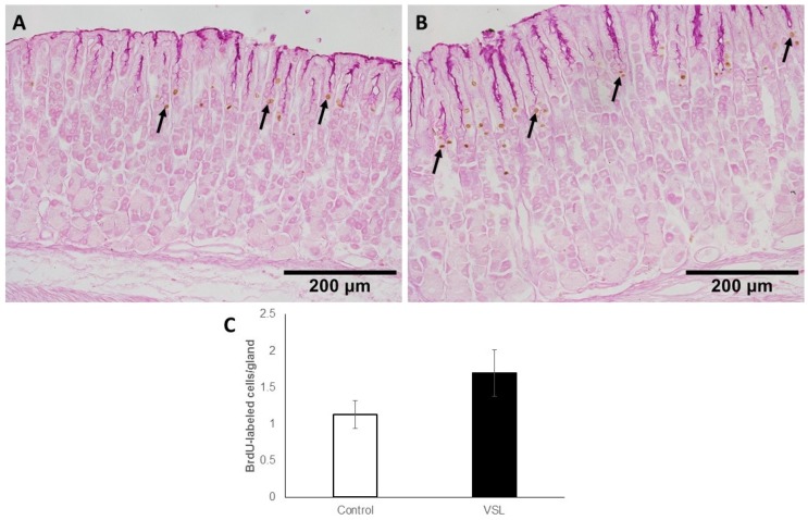
Figure 9.
Immunohistochemical analysis of S-phase cells using anti-5′-bromo-2′-deoxyuridine (BrdU) antibody in the gastric mucosa of control (A) and VSL-treated (B) mice. Note that BrdU-labelled cells (brown nuclei) of VSL-treated tissues tend to appear more expanded when compared with control tissues and uniformly distributed in most of the glands. Magnification bar = 200 µm. Analysis of BrdU-labeled cell counts per gland in the mucosa of seven control and seven VSL-treated mice are presented as mean ± SE (C). No significant difference was found between VSL and control tissues.

