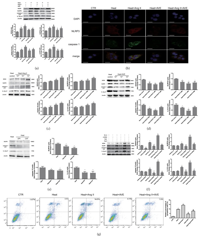
Figure 6.
AVE 0991 attenuated pyroptosis and hepatocyte damage induced by heat stress by inhibiting the ROS-NLRP3 inflammatory signalling pathway in vitro. (a) The protein expression levels of NOX4, NLRP3, caspase-1 and IL-1β in BRL -3A cells were analysed by western blotting. ∗P<0.05 compared with the control group. #P<0.05 compared with the heat group. &P<0.05 compared with the heat+Ang II group. (b) Immunofluorescence staining was conducted and observed using confocal laser microscopy. The scale bar indicates 20 μm. NLRP3: red; caspase-1: green. The nuclei stained with DAPI (blue). (c, d) BRL-3A cells subject to heat stress were treated with varying concentrations of Ang II and AVE 0991. The quantified relative protein expression analysed by western blotting is shown in the graph (right). ∗P<0.05 compared with the heat group. (e) The effect of 10-5 M DPI and 10 mM CAT on BRL-3A cells pre-treated with heat stress. ∗P < 0.05 compared with the heat group as a control. (f) Protein expression after siRNA transfection targeting NOX4. ∗P<0.05 compared with the control group. #P<0.05 compared with the heat group. △P<0.05 compared with the heat+Ang II group. (g) The expression of activated caspase-1 was analysed by flow cytometry. ∗P<0.05 compared with the sham or control groups. #P<0.05 compared with the heat group. &P<0.05 compared with the heat+Ang II group. n=6 per group. The data are presented as the mean ± SD. All of the assays were performed in triplicate. DPI: diphenylene iodonium; CAT: catalase.

