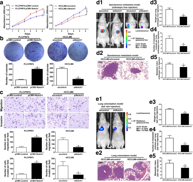
Fig. 2.
Notch1 promotes metastasis in HCC. Proliferation was examined by Cell Counting Kit-8 (CCK-8) (a) and colony formation assay (b). Migratory and invasive ability of Notch1-modulated cells were examined by Transwell assays (c). (d) In vivo spontaneous metastasis assay. HCC cells were injected into the left hepatic lobe of nude mice. (d1) Bioluminescence imaging of spontaneous metastasis was taken 10 weeks after orthotopic implantation. Right panel: bioluminescence imaging of metastasis taken after masking of the signal from primary xenografts. (d2) Lung metastasis was confirmed by H&E staining. (d3) Lung weights and (d4) number of lung metastatic nodules in each group. (d5) The metastasis index was calculated from the number of nodules/lung weight ratio. e In vivo lung colonization assays. The indicated stable cells were injected to nude mice via tail vein. (e1) Bioluminescence imaging and (e2) H&E staining of the lung metastatic tumors, respectively. (e3) Lung metastatic nodules, (e4) weights and (e5) metastasis index of nude mice in each group. *: P < 0.05

