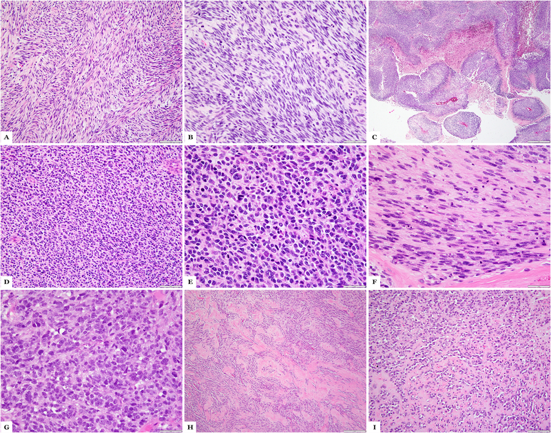
Figure 2:
MYOD1-mutant rhabdomyosarcoma with primitive round to spindle cell component. (A-C) Case 1 showing areas of classic spindle cell morphology with fascicular growth (A), solid zones of primitive appearing spindle cell component (B) and geographic necrosis (MPNST-like); (D-F) Case 28 showing tumor in the liver with undifferentiated primitive round cell component (D, E) and areas of spindling (F); (G-I) Case 23, a post-therapy resection, showing primitive round cell areas (G), spindle cell morphology (H) and areas with sclerosing morphology (I). (H&E stains)

