
Figure 1. Exosomes containing small RNAs are secreted into the medium of fetal SMGs cultured ex vivo.
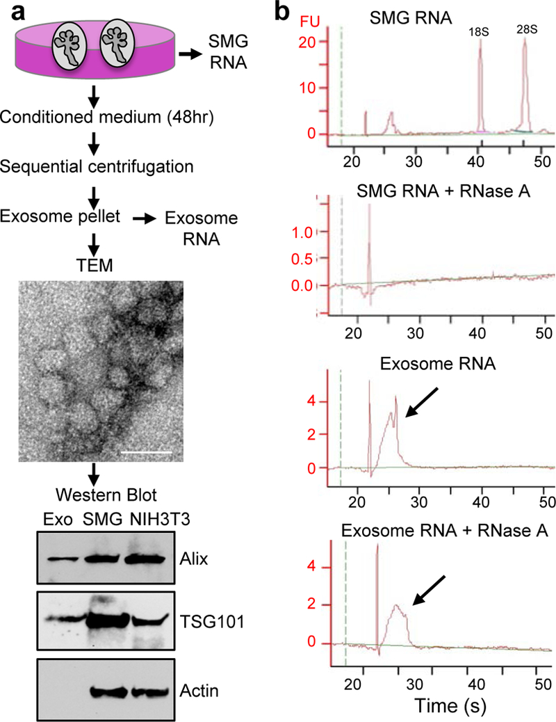
(a) Exosomes (Exo) isolated by sequential centrifugation were analyzed by transmission electron microscopy (TEM, Scale bar = 50 nm) and Western blot for the exosome markers Alix and TSG101. Intact SMG and NIH3T3 cell lysates are positive controls. (b) Bioanalyzer analysis shows that small RNAs in exosomes were resistant to RNase A degradation. Arrows indicate the peak of small RNA before and after RNase A treatment. FU; fluorescence units. N = 3, graph is a representative experiment.