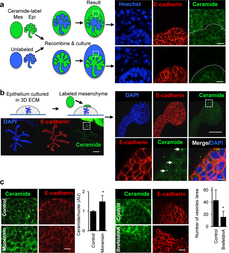
Figure 2. Fluorescently-labeled exosomes are transported from the SMG mesenchyme to the epithelium.

(a) SMG epithelium (Epi) separated from mesenchyme (Mes) were both labeled with fluorescent ceramide and recombined with unlabeled epithelium or mesenchyme. After 12 h of recombination they were analyzed by confocal microscopy. The ceramide label was transported from the mesenchyme to the epithelium but not the other way around. Scale bar = 10 μm. (b) Isolated epithelium in 3D laminin extracellular matrix was cultured with labeled mesenchyme. Labeled vesicles were detected in the epithelium after 8 h (white arrows). Scale bar = 200 μm (left), 50 μm (right upper), 5 μm (right lower). (c) Monensin (1 μM) increases ceramide-labeled vesicles in the epithelium, whereas brefeldin-A (1 μg/ml) decreases the number of vesicles in the epithelium. Scale bars = 10 μm. Student’s t-test, * P< 0.05.
