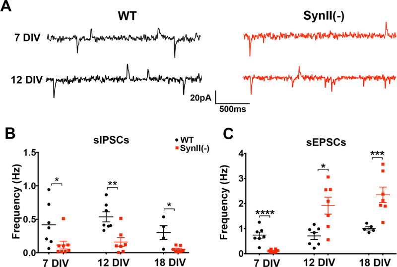
Fig. 1.
Excitatory and inhibitory spontaneous transmission in SynII deleted neurons at different developmental stages in vitro. A. Representative recordings showing sEPSCs (downward polarity) and sIPSCs (upward polarity) recorded at – 15 mV holding potential. B. Spontaneous inhibitory transmission is uniformly reduced in SynII(−) neurons. C. In SynII(−) neurons, spontaneous excitatory transmission is reduced at the initial developmental stage (7 DIV) but enhanced at subsequent developmental stages (12 and18 DIV). * p<0.05; * <0.01; *** p<0.005, **** p<0.001.

