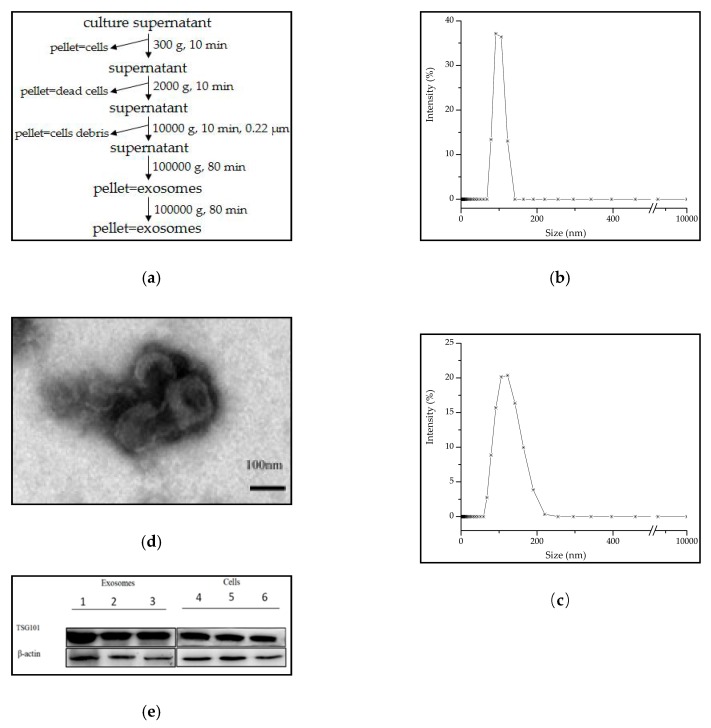
Figure 1.
Exosomes isolation and characterization. (a) The isolation method of the culture cell-derived exosomes; (b) DLS result showed the size of MCF7 cell-derived exosomes about 100 nm in mode; (c) and EC109 cell-derived exosomes about 120 nm; (d) TEM displayed the morphology and size of exosomes which were negatively stained; (e) Detection of TSG101 expression on cell derived exosomes (1–3) and its original cells (4–6), β-actin as control. Note: 1. MCF7-exosome; 2. M231-exosome; 3. HepG2-exosome; 4. MCF7 cell; 5. M231 cell; 6. HepG2 cell.

