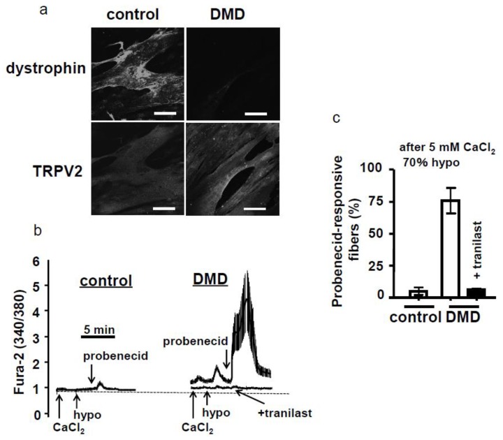
Figure 4.
Characterization of the control and human Duchenne muscular dystrophy (DMD) myotubes and effects of tranilast on the DMD myotubes. (a) Human control myotubes (KD3) and dystrophic myotubes (D4P4) produced from human DMD patients were visualized by immunofluorescence using an anti-TRPV2 or anti-dystrophin antibody. Scale bar = 50 μm. (b) Representative traces for the intracellular Ca2+ response. Myotubes placed in a solution containing 2 mM CaCl2 were stimulated with high Ca2+ (5 mM CaCl2, indicated by arrow) and then a 70% hypoosmotic medium (hypo, indicated by arrow). Myotubes were further perfused with the medium containing 38 μM probenecid (probenecid, indicated by arrows). In one experiment, 100 μM tranilast was included in the perfusion medium. (c) The ΔF-ratio was calculated by subtracting the resting fluorescence ratio from the maximal ratio after the inclusion of probenecid. Myotubes exhibiting a ΔF-ratio > 0.3 were defined as probenecid-responsive myotubes. Only DMD myotubes were responsive to probenecid, which was blocked completely by tranilast [34].

