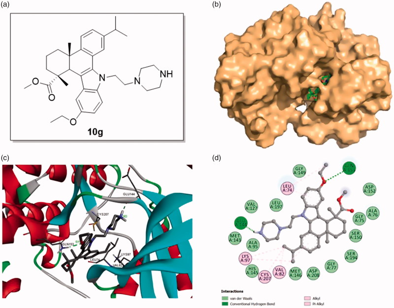
Figure 4.
Binding mode of compound 10g at MEK1 kinase domain (PDB: 3EQF). (a) Molecular structure of compound 10g; (b) Space filling model of MEK1 protein with compound 10g embedded in the binding pocket; (c) Binding pose of compound 10g within the MEK1 kinase domain. Ligand and key residues are presented as stick models and colored by atom type, whereas the proteins are represented as ribbons. The dash lines exhibit the hydrogen bond interactions; (d) 2D projection drawing of compound 10g docked into MEK1 active site.

