Fig. 3.
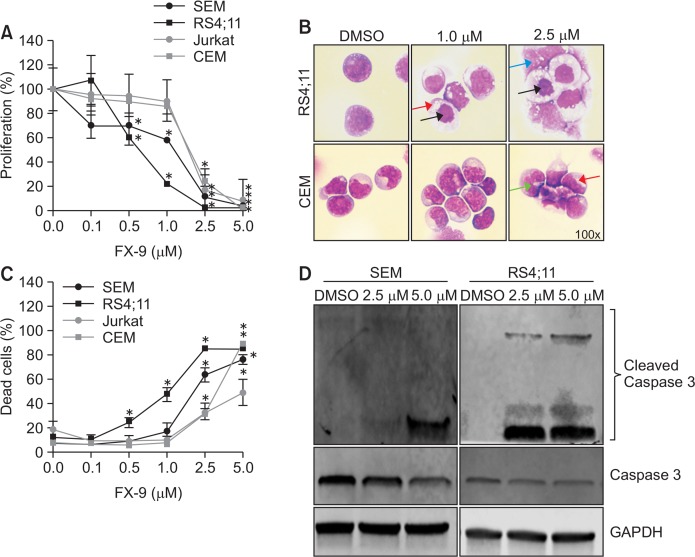
FX-9 inhibits cell proliferation and induces changes on ALL cell morphology. B- and T-ALL cells were exposed to FX-9 and DMSO for up to 72 h to evaluate cell proliferation, morphology and cell death. (A) FX-9 significantly inhibited cell proliferation of B- and T-ALL cells after 72 h. (B) Cellular morphology was assessed by May-Grünwald staining. Results are exemplary displayed for RS4;11 and CEM after 48 h. Cytomorphology was altered in both cell lines. FX-9 treatment induced an extensive cytoplasmic vacuolization (red arrow), karyopyknosis (black arrow), karyorrhexis (green arrow) and karyolysis (blue arrow). (C) The amount of apoptotic and necrotic cells (dead cells) in B- and T-ALL cell lines was analyzed using annexin V and propidium iodid staining after 72 h. FX-9 significantly induced apoptosis in all cell lines. (D) Western blot revealed that apoptosis was induced via caspase 3 pathway as indicated by an increased amount of cleaved caspase 3 protein in FX-9 treated SEM and RS4;11 cells after 48 h. Results are displayed as mean ± standard deviation of three independent experiments. Asterisk represents a statistically significance difference between FX-9 and DMSO treated cells with a *p<0.05.

