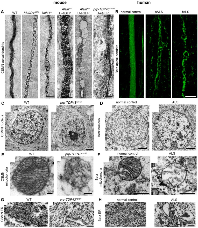
Figure 2.
Betz cell pathology is similar between mouse and human at a cellular level. (A) Apical dendrites of CSMN displaying vacuoles in brains of various mouse models of motor neuron disease. (B) Apical dendrites of Betz cells displaying vacuoles in brains of patients with sporadic or familial ALS. (C–H) Electron microscope images showing pathology of various organelles in CSMN of prp-TDP43A315T mouse and Betz cells of ALS patients. Observe similar nuclear membrane defects (C,D), mitochondria defects (E,F), and endoplasmic reticulum defects (G,H). Scale bars: 5 μm (brightfield, left), and 1 μm (E.M., right) in (A), 10 μm in (B), 2 μm in (C), 500 nm in (D), 200 nm in (E), 500 nm in (F), 1 μm in (G–H).

