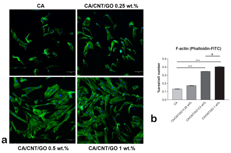
Figure 3.
Cytoskeleton developed by human adipose-derived stem cells (hASCs) cultivated in contact with CA-CNT-GO materials, scale bar 100µm. (a) Fluorescence images of the actin filaments (green) and nuclei (blue) of hASCs and (b) quantification of the phalloidin-FITC staining normalized to cell number. Statistical significance: # p < 0.05; *** p < 0.001.

