Figure 5.
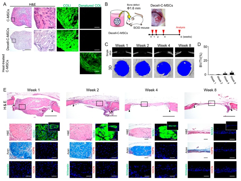
Transplantation of Decell-C-MSCs failed to induce bone regeneration. (A) C-MSCs, Decell-C-MSCs, and heat-treated C-MSCs were prepared as described in the Materials and Methods section. Semi-serial sections were stained with H&E and immunostained with anti-COLI antibody and 5-FAM conjugated-collagen hybridizing peptide, as indicated. Left panels show lower magnification. Bar = 200 µm. The second to fourth panels are magnifications of the boxed regions. Bar = 50 µm. (B) Study design for the in vivo experiment. Decell-C-MSCs were transplanted into a SCID mouse calvarial defect 1.6 mm in diameter with no artificial scaffold. (C) Representative µCT images at 1, 2, 4, and 8 weeks after surgery. (D) Ratio of the segmented bone volume (BV) to the total volume (TV) of the defect region at 1, 2, 4, and 8 weeks following surgery. Data are mean ± SD of four mice per group. (E) Animals were sacrificed at 1, 2, 4, and 8 weeks after surgery and the calvarial bones were fixed. Semi-serial sections were obtained and stained with H&E and AZAN and immunostained with anti-human vimentin, anti-human COLI, anti-human OPN, and anti-human OCN antibodies, as indicated. Nuclei were counterstained with DAPI for immunostaining. Upper panels show lower magnification. Bar = 200 µm. Bottom panels are magnifications of the boxed region. Bar = 50 µm. White boxes indicate human COL1 expression in the whole defect area. The photographs are representative of four independent experiments.

