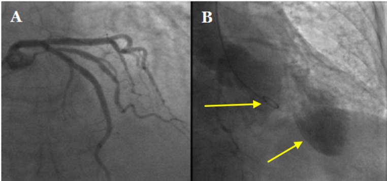
Figure 2.
(A) Coronary angiogram showing smooth, obstructed left main stem, left anterior descending and circumflex arteries. (B) Left ventriculogram showing apical ballooning in keeping with Takotsubo cardiomyopathy. The arrows delineate the 5F pigtail catheter within the basal portion of the left ventricular cavity and the ballooning appearance in the left ventricular apex.

