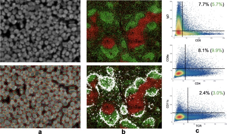
Fig. 11.
BALB/c spleen (slide 1) CODEX image segmentation/quantification results. a ~ 200 cells with DRAQ5 nuclear stain (above) and corresponding cell/nucleus segmentation (below). b ~ 71 k cells in stitched, downscaled 9072 × 9408 pixel image showing IgD (green) and CD90 (red) expression as well as location of CD169+ marginal zone macrophages as white dots. c Double positive cell population rates post cleanup-gating with expected percentages in green

