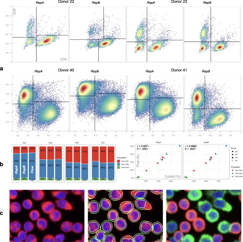
Fig. 8.
T cell population recovery comparison (notebook). a CD4/CD8 gating results, as determined by automatic gating functions in OpenCyto [25], over two imaging replicates for each of 4 donors. While all images were collected over a field of view of the same size, samples for donors 40 and 41 were prepared at 3x higher cell concentrations to demonstrate that segmentation and intensity measurements are robust to greater image object densities. b Cell population size for both replicates compared to a single flow cytometry measurement for each donor as well as Pearson correlation demonstrating strong agreement between the two (r > 0.99, P < 0.0001, two-tailed t-test). c Cell images from donor 41 showing (from left to right): DAPI (blue) and PHA (red) stain, DAPI and PHA with cell and nuclei segmentations, and DAPI with CD4 (red) as well as CD8 (green). See supplementary file Additional file 1: Figure S1 for a comparison of these results to those from the same workflow without much of the gating used here to remove invalid cells

