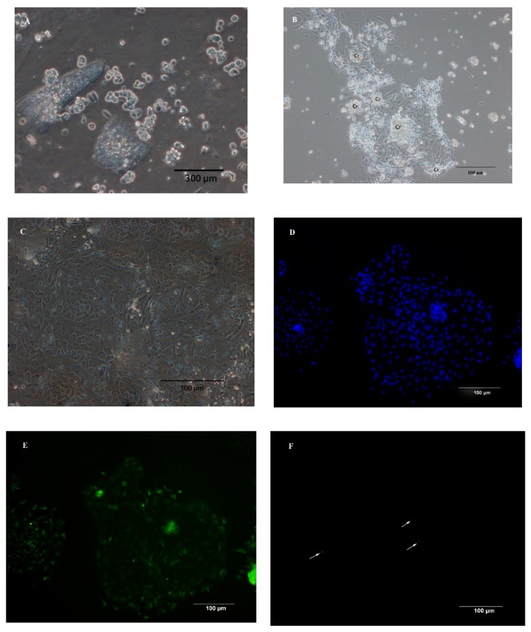
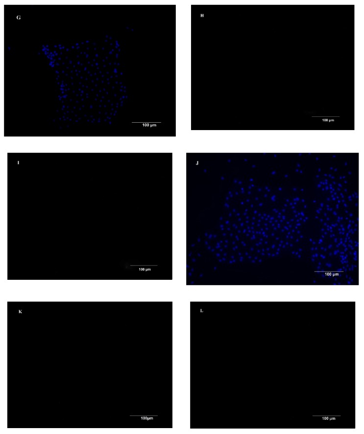
Figure 1.
Morphological features and immunofluorescence staining of primary chicken intestinal epithelial cells (IECs). (A) Epithelial cells were isolated in cell clusters. (B) Primary chicken IECs at 24 h post-seeding. The image shows cobblestone-shaped IECs radially growing from the center of the crypt (Cr) and cell clusters connecting. (C) Primary chicken IECs culture within 4–5 days post-seeding. Image shows a confluent chicken IECs. (D,G,J) Nuclei were stained (blue). (E) Immunostaining against cytokeratin-18 (green) at 3 days post-seeding. (F) Immunostaining against vimentin (red, the arrows in the figure) at 3 days post-seeding. (H) Primary chicken IECs presented in the image (G) were incubated only with the primary antibody of cytokeratin without the secondary antibody to serve as a primary antibody control. (I) Primary chicken IECs presented in the image (G) were incubated first with buffer only without the primary antibody of cytokeratin-18 and subsequently with secondary antibody to serve as a secondary antibody control. (K) Primary chicken IECs presented in the image (J) were incubated only with the primary antibody of cytokeratin without the secondary antibody to serve as a primary antibody control. (L) Primary chicken IECs presented in the image (J) were incubated first with buffer only without the primary antibody of cytokeratin-18 and subsequently with secondary antibody to serve as a secondary antibody control.


