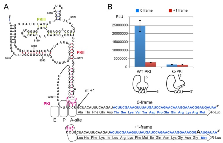
Figure 4.
Translation of R-Luc driven by the CrPV IGR and analysis of potential +1-frame translation. (A) The secondary structure of the IGR IRES. The IGR is driven translation of Renilla luciferase (R-Luc) fused in the 0-frame or +1-frame. To monitor the +1-frameshifting activity, one extra A nucleotide was added in the R-Luc sequence. Nucleotides and amino acid residues in blue are from R-Luc. The pseudoknot PKI which mimics the codon/anticodon is drawn in pink. The A-, P- and E-site of the ribosome are schematized. Nucleotide numbering is from the CrPV genome. (B) Histogram representing the luciferase activities in 0-frame or +1-frame starting from the WT or knockout PKI. Transcripts were synthesized in vitro, denatured/renatured and incubated in Rabbit Reticulocyte Lysates at 30 °C for 2 h. Translation of R-Luc was measured by measuring the bioluminescence produced with a Dual-Glo luciferase assay (Promega) in a luminometer. Translation efficiencies are represented as raw bioluminescence activity (Relative Light Units or RLU) and resulted from at least 3 independent experiments. Standard deviations are shown.

