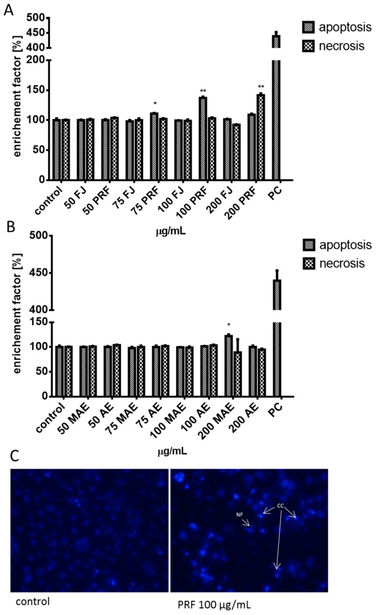
Figure 10.
The influence of 24 h of exposure of juice (FJ) and PRF (phenolic rich fraction) (A), MAE (methanol-acetone extract) and AE (acetone extract) (B) on DNA fragmentation (late stage of apoptosis) analyzed by Cell Death Detection kit, PC—internal positive control of assay; nuclear morphology of DAPI-stained Caco-2 cells after 24 h of exposure to PRF at 100 μg/mL dosage. NF (nuclear fragmentation) and CC (chromatin condensation) in cells were observed (C) using fluorescence microscope (Nikon TS100 Eclipse, Japan), 200× magnification. Images are representative of one of four similar experiments. Control cells were not exposed to any compound but the vehicle; values are means ± standard deviations from at least three independent experiments; statistical significance was calculated versus control cells (untreated) * p ≤ 0.05, ** p ≤ 0.01.

