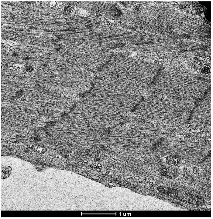
Figure 2.
Transmission electron microscopy image of cardiomyocytes produced from fibroblast-derived iPSCs. The reprogramming technology was used to convert cultured human fibroblasts into iPSCs and then into cardiomyocytes (Cell Applications, Inc., San Diego, CA, USA). Cells were then fixed in the 2.5% glutaraldehyde/0.05 M cacodylate solution, post-fixed with 1% osmium tetroxide and embedded in EmBed812 (Electron Microscopy Sciences Hatfield, PA, USA). Ultrathin sections (70 nm) were post-stained with uranyl acetate and lead citrate and examined in the Talos F200X FEG transmission electron microscope (FEI, Hillsboro, OR, USA) [18]. The image shows the cells developed from fibroblasts having clear striations and sarcomere structures. Magnification 5500×.

