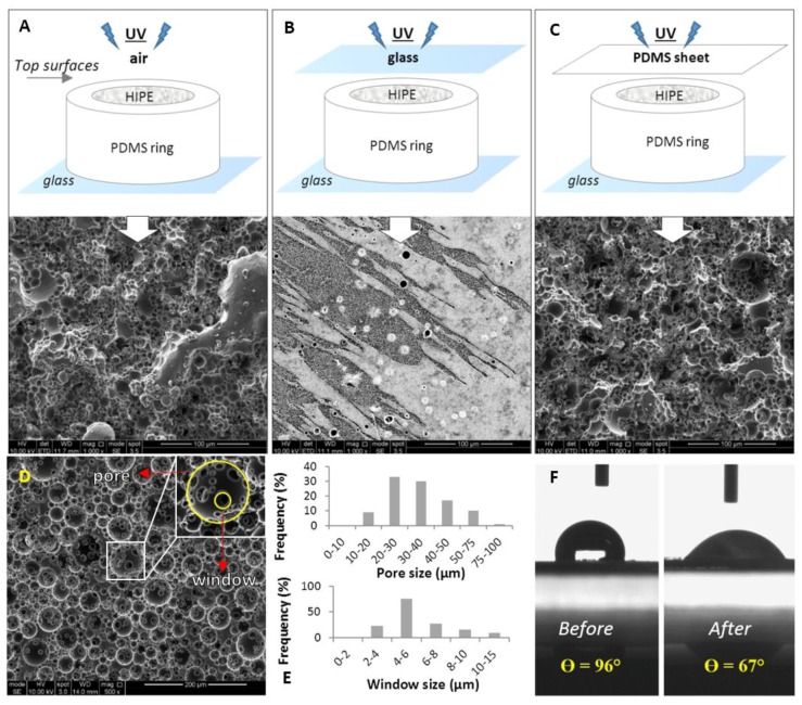
Figure 2.
Scanning electron microscope (SEM) images of the top surfaces PCL polyHIPEs cured in contact with; (A) air, (B) glass, and (C) polydimethylsiloxane (PDMS) sheet. (D) SEM image of the transverse section of PCL polyHIPEs. (E) Pore size and window size distributions of the inner section. (F) Contact angle measurements of a water droplet on PCL polyHIPE before and after air plasma treatment (n = 3).

