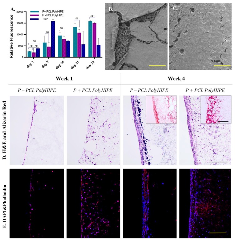
Figure 3.
(A) Metabolic activity of murine long-bone osteocytes (MLO-A5s) cultured on P-, P+ PCL polyHIPEs, and tissue culture plate (TCP) for 4 weeks. SEM images of the top surfaces of (B) P+ and (C) P- PCL polyHIPEs cultured MLO-A5s on for 4 weeks (Scale bar represents 500 µm). (D) Haematoxylin and eosin (H&E) and Alizarin red, and (E) fluorescent staining of MLO-A5s cultured on P+ and P- PCL polyHIPEs for 1 week and 4 weeks (Scale bar represents 250 µm, blue: 4’,6-diamidino-2-phenylindole (DAPI), red: phalloidin tetramethylrhodamine (TRITC)).

