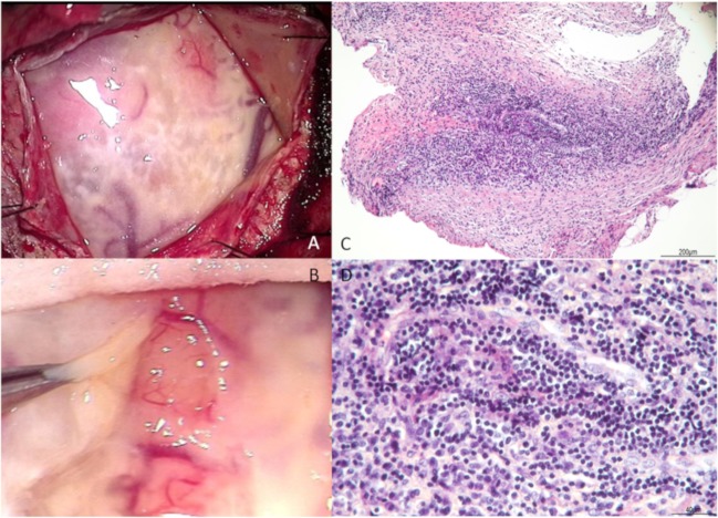
Figure 3.
During the meningeal biopsy, there were lots of viscous pale yellow effusions in the subdural (A) and the subarachnoid (B). Histopathology (H&E stain) of the meningeal biopsy: a low-power micrograph of the leptomeninges showed fibrous thickening changes (C, ×10) and a high-power micrograph of the area revealed infiltration with plasma cells and lymphocytes (D, ×40).

