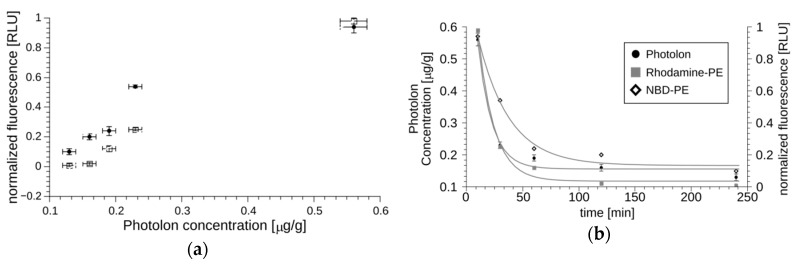
Figure 4.
The pharmacokinetic characteristics of liposomal Photolon injected in the jugular vein of Sus scrofa f. domestica. Panel (a) shows the correlation between the fluorescence of Photolon and NBD-PE or Rhodamine-PE measured in blood. All points in the plot were determined for a specific time-point, for which both the fluorescence of entrapped Photolon and the labeled lipid capsule were quantitated, and the resulting values are shown on the OX and OY axis, respectively. The experimental data can be fitted with straight lines, which indicates that Photolon remains in liposomes throughout the duration of the experiment. Panel (b) shows a pharmacokinetic curve for fluorescently labeled lipids and Photolon. The decreasing fluorescence values (with half-time equal to approximately 20–30 min indicating efficient elimination).

