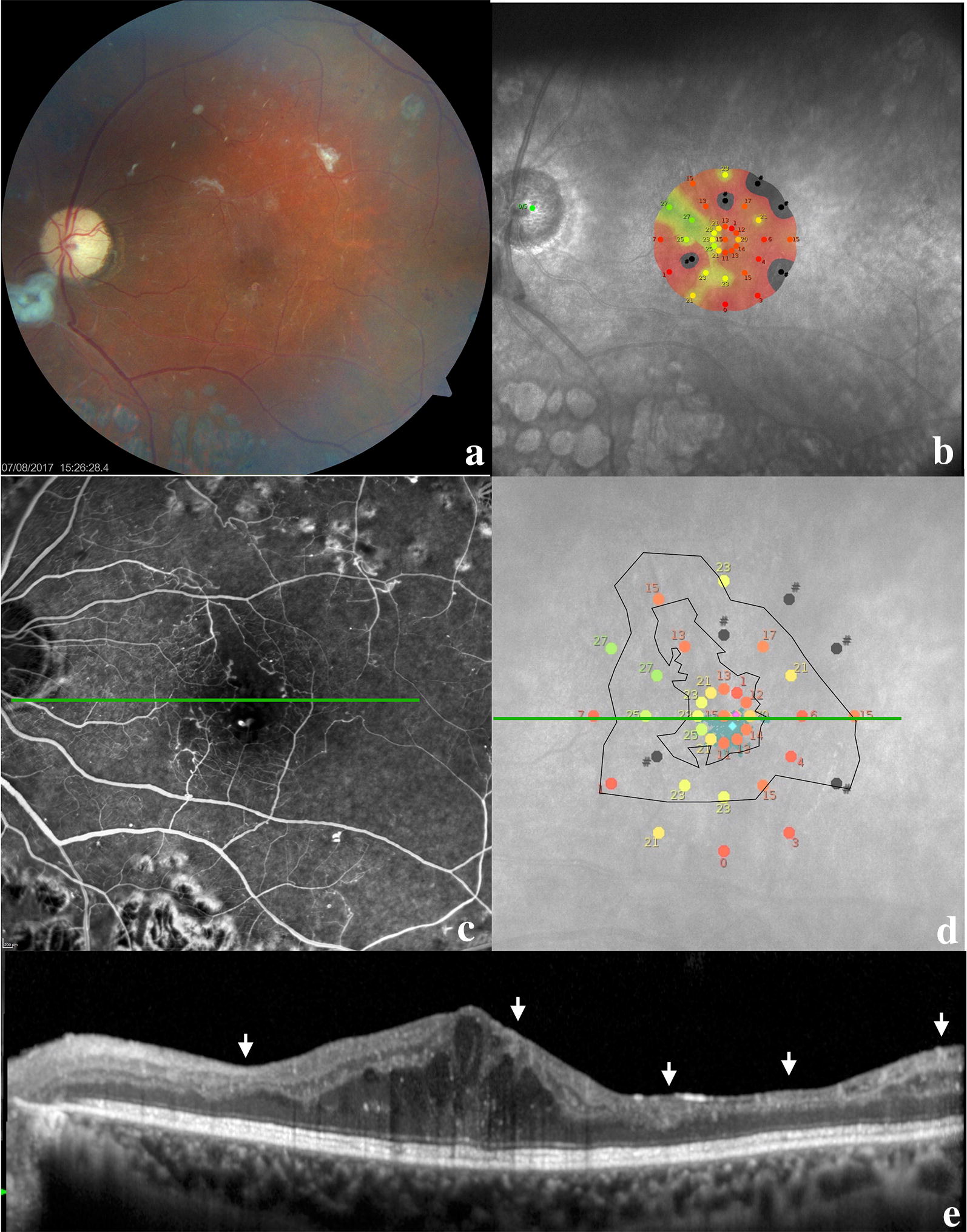
Fig. 2.

Multimodal image of a patient at 6 months follow-up. a Color retinography with pallor of the optic disc, macula with edema and fibrotic tissue, small hemorrhagic dots in the posterior pole, and panretinal photocoagulation beyond the temporal arcades; b microperimetry demonstrating macular sensitivity; c fluorescein angiography in the venous phase demonstrating a large foveal avascular zone (FAZ) with capillary dropout at the border of the FAZ; d macular sensitivity superimposed on the FAZ area (smaller black circle) and the area with some degree of hypoperfusion (larger black circle); e this OCT line represents the green line in the middle images; the image demonstrates the persistence of edema and the disorganization of retinal inner layers (DRIL) as indicated by the white arrows; these locations correlate with worse macular sensitivity on microperimetry and capillary dropout and hypoperfusion on fluorescein angiography
