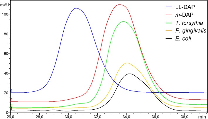
Fig. 4.
Separation of m-DAP and LL-DAP by reversed-phase HPLC after dabsylation, revealing the preponderance of m-DAP in all analysed peptidoglycan isolates. Overlay of chromatograms for T. forsythia peptidoglycan (green line), P. gingivalis peptidoglycan (yellow line) and E. coli peptidoglycan (black line) and the standards m-DAP (red line) and LL-DAP (blue line)

