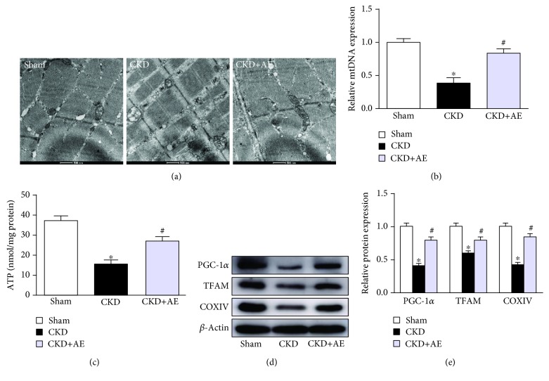
Figure 6.
Aerobic exercise attenuated muscle mitochondrial dysfunction in CKD mice. (a) Morphological changes of mitochondria examined by transmission electronic micrographs (TEM) (magnification ×12,000). (b) Mitochondrial DNA (mtDNA) copy number, normalized against GAPDH, measured via real-time PCR. (c) ATP production, expressed as per mg protein. (d) Western bolt of PGC-1α, TFAM, and CoxIV expression. (e) Quantification analysis of PGC-1α, TFAM, and CoxIV levels, normalized against β-actin. ∗P < 0.05, the Sham group vs. the CKD group. #P < 0.05, the CKD+AE group vs. the CKD group. Data represent the mean ± SEM (n = 6).

