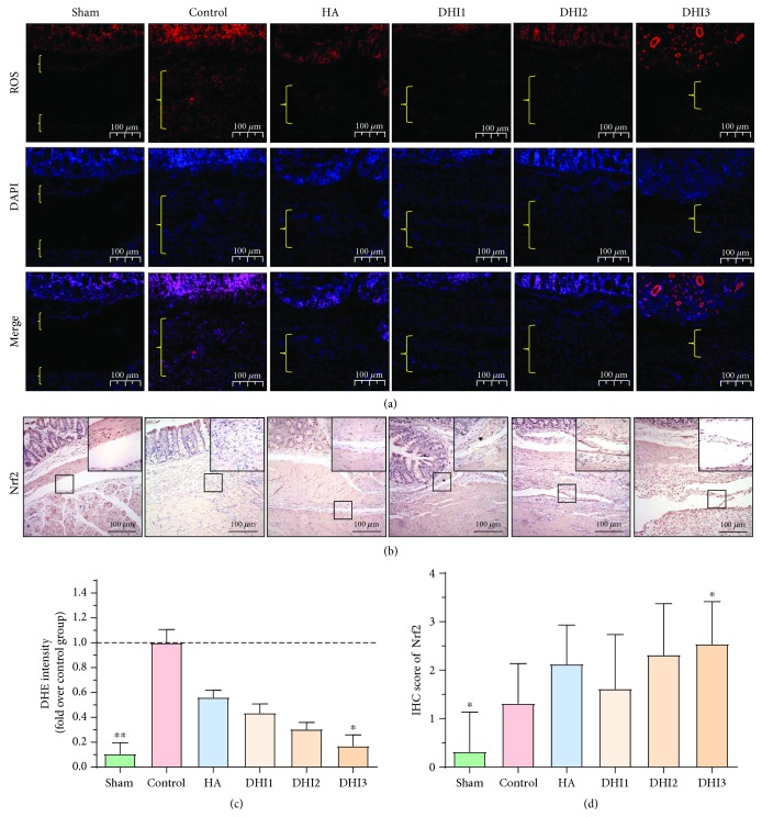
Figure 5.
DHI treatment reduces oxidative stress in the adhesion tissues of the rat model 7 days after operation (n = 8; compared with the control group, ∗P < 0.05). (a) Representative ROS level of each group at 100x magnification, and the area between the bracket indicates the tissue in the adhesion area. (b) Nrf2 staining scores of each group at 100x magnification. The top right corner is shown at 200x magnification. (c) Relative ROS expression in the adhesion tissue of each group (compared with the control group, ∗P < 0.05, abnormal distribution, Kruskal-Wallis test). (d) Nrf2 staining scores of each group (compared with the control group, ∗P < 0.05, normal distribution, one-way ANOVA).

