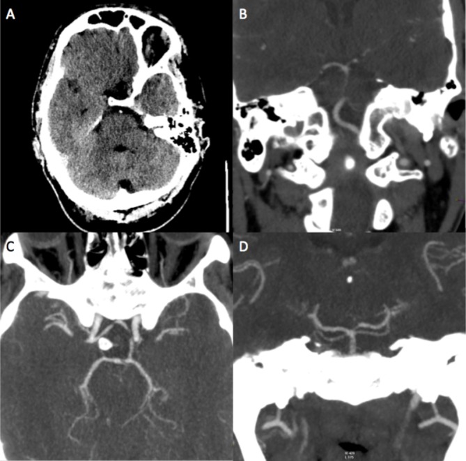
Figure 1.

Non-contrast CT demonstrates a hyperdense basilar artery in keeping with acute thrombosis (A). Enhanced CT angiogram confirms basilar artery occlusion as well as posterior inferior cerebellar artery termination of the right vertebral artery (B). Posterior circulation was partly supplied via a patent left posterior communicating artery (C); however, the P1 segment of the left posterior cerebral artery was congenitally hypoplastic (D) meaning it was not a feasible route for thrombectomy.
