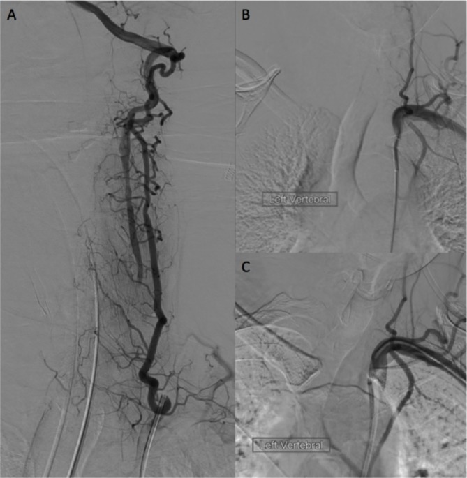
Figure 2.

Digital subtraction angiography following injection of the left thyrocervical trunk (A) reveals distal filling of the left vertebral artery (VA) via collaterals. Despite attempts from the groin (B) and radial access sheath (C) the occluded left VA origin could not be accessed.
