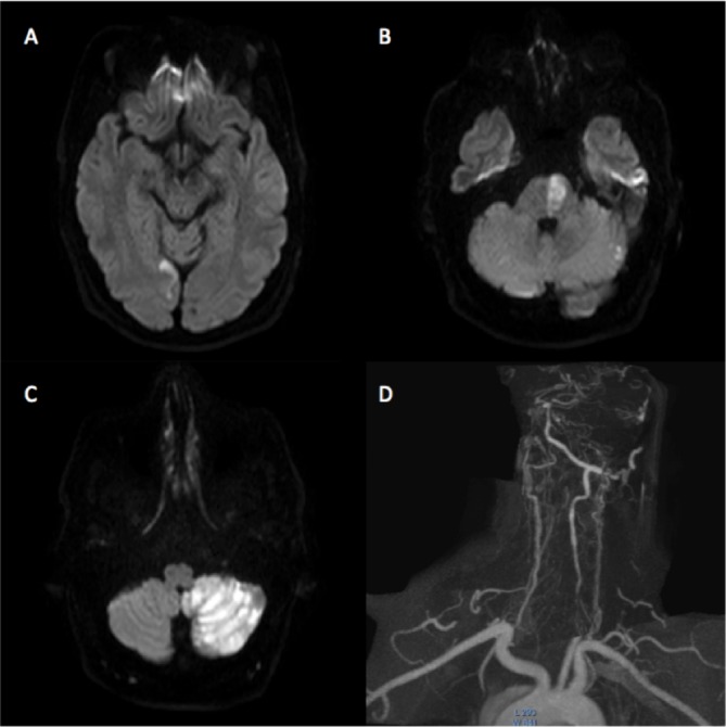
Figure 4.

Diffusion weighted MRI sequences performed at day 1 postprocedure demonstrate areas of infarction with the medial right occipital lobe (A), left hemipons (B) and left cerebellar hemisphere (C). MR angiography confirms persistent occlusion of the left vertebral artery origin, however there is distal filling via collaterals (D).
