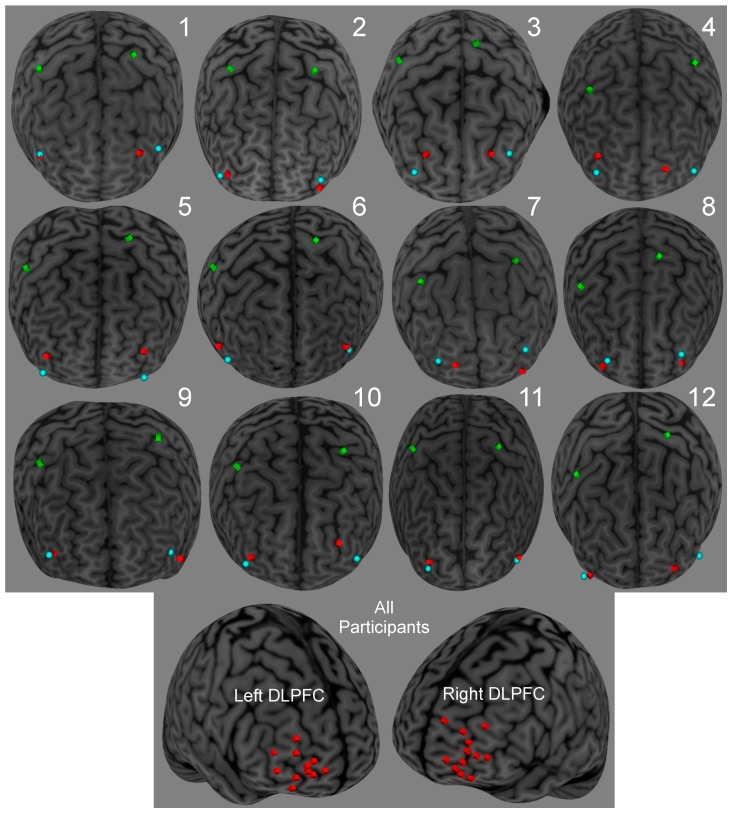
Figure 2.
Dorsolateral prefrontal cortex (DLPFC) anatomical, DLPFC stimulation and M1 stimulation locations for Experiments 1–4 for the 12 participants based on individualized MRIs using neuronavigation software. We adjusted the targeted DLPFC stimulation sites to accommodate for both transcranial magnetic (TMS) coils on the scalp. Green cubes denote M1 stimulation locations, blue spheres denote DLPFC anatomical locations, and red cubes denote DLPFC stimulation locations.

