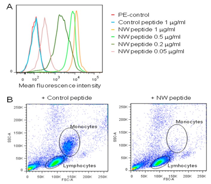
Figure 2.
Binding and depletion of blood monocytes. (A) Representative flow cytometry histograms showing the peptide binding to purified blood monocytes. Cells were incubated with various concentrations of the biotinylated NW peptide, followed by streptavidin-conjugate PE and analysis by flow cytometry. (B) Monocyte depletion. PBMCs were incubated with the biotinylated NW peptide or control peptide (20 μg/mL each), and then processed as described in Material and Methods. The cells were analyzed by flow cytometry to check for monocyte depletion after peptide addition and capture on streptavidin beads. The data are representative of four independent experiments.

