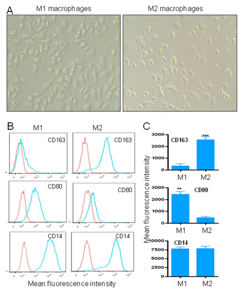
Figure 4.
Monocyte-derived M1 and M2 macrophages. (A) Morphology of monocyte-derived M1 or M2 macrophages in X-vivo 15 medium. Original magnification, x20. (B) Phenotypic characterization of M1 and M2 macrophages. Cell surface expression of CD163, CD80 and CD14 markers by M1 and M2 macrophages. (C) Quantitative data from three independent experiments are presented as a mean ± SD. ** p < 0.01, *** p < 0.001.

