Figure 5.
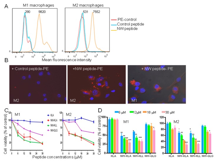
Effects of the fusion lytic peptides on M1 and M2 macrophages. (A) Representative flow cytometry histograms showing the binding of the NW peptide to M1 and M2 macrophages. The numbers indicate the mean fluorescence intensities of the peptide binding. (B) Internalization of the NW-peptide-streptavidin-PE complexes by M2 and M1 macrophages. Biotinylated NW peptide- or control peptide-streptavidin-PE complexes were added to M2 macrophages and incubated for 40 min at 4 °C. After washing, the cells were incubated at 37 °C for 60 min, further washed, fixed, and then confocal microscopy images were taken. Original magnification, x40. The uptake of the NW peptide-PE complexes by M1 is also shown. (C) Cell viability. The cells were incubated with various concentrations of the tested peptides for 60 min at 37 °C, and cell viability was determined using the CellTiter 96R Aqueous One Solution reagent. The results are represented as mean ± SD of triplicate determination. Quantitative data (mean ± SD) from three independent experiments are shown in (D). ** p < 0.01, *** p < 0.001.

