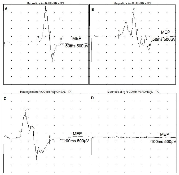
Figure 1.
Examples of MEPs recordings in MPS patients. (A) normal MEP from the upper limb; (B) MEP with reduced amplitude and polyphasic shape from the upper limb; (C) normal MEP from the lower limb; (D) absence of transcranially-induced motor response from the lower limb. MPS = mucopolysaccharidosis; Magnetic stim = transcranial magnetic stimulation; R ULNAR = right ulnar nerve; R COMM PERONEAL = right common peroneal nerve; FDI = first dorsal interosseous muscle; TA = tibialis anterior muscle; MEP = motor evoked potential; 50 ms = temporal resolution of the screen (sweep) for upper limb recordings; 100 ms = temporal resolution of the screen (sweep) for lower limb recordings; 500 µV = amplification factor of the screen.

