Figure 1.

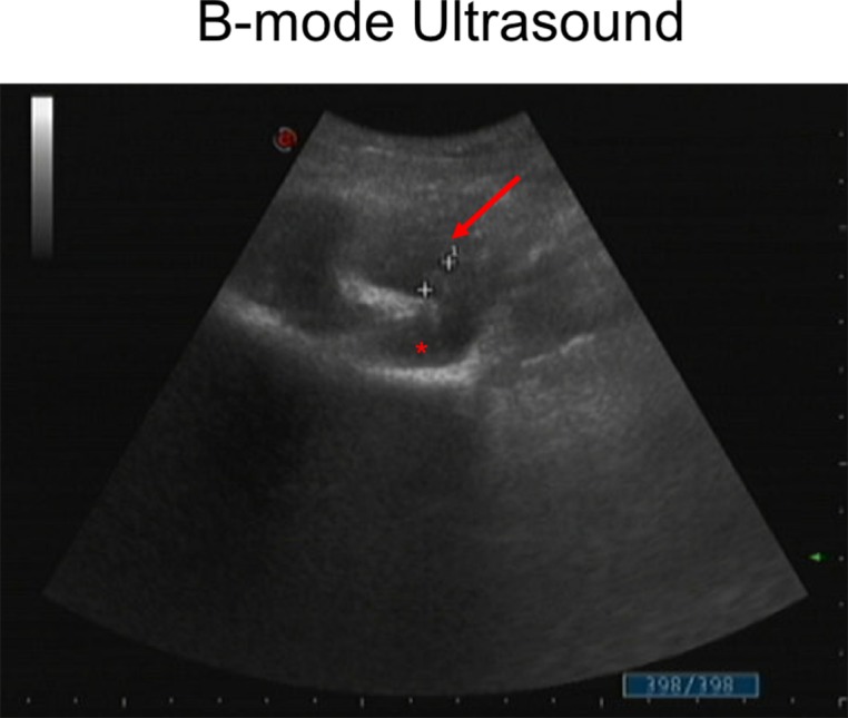
B mode ultrasound images of acute appendicitis (arrow) demonstrating focal, oedematous wall thickening with a maximum appendix diameter of 7.6 mm (white crosses) and surrounding fluid accumulation (asterisk).

B mode ultrasound images of acute appendicitis (arrow) demonstrating focal, oedematous wall thickening with a maximum appendix diameter of 7.6 mm (white crosses) and surrounding fluid accumulation (asterisk).